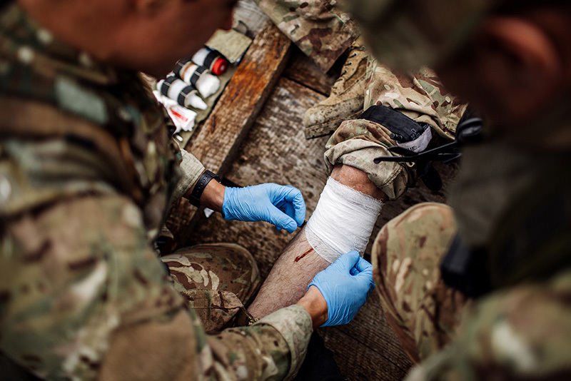
In Iraq in 2007 five hundred pounds of explosives detonated beneath Combat Medic Greg Dotson’s Bradley Fighting Vehicle. His unit soldiers, trained by Greg, pulled him and his medic bag from the wreck and began work. While a furious firefight was going on around them, they applied tourniquets high on his shredded legs, slid a breathing tube down his nose and throat and got a field dressing secured tightly around the shrapnel wounds in his abdomen. They tried to start an IV, but his veins were flat. Splints were taped to his legs and they strapped his 6”6” frame to a stretcher and a Blackhawk medevac helicopter picked him up. Minutes later he was on the operating room table at the closest U.S. air base.
|
|
|
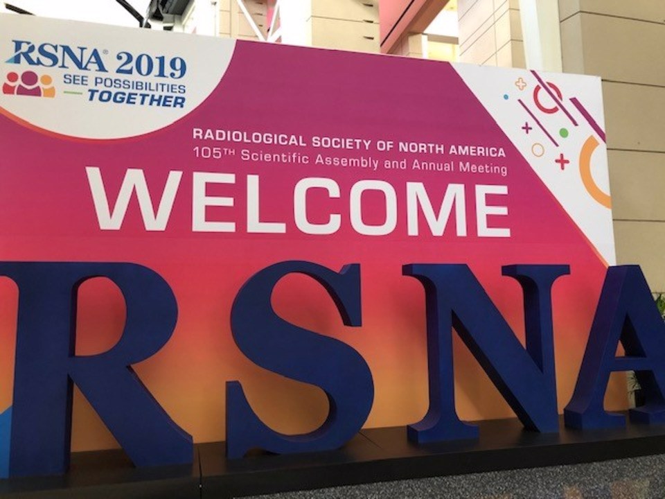
Welcome to RSNA 2019: See Possibilities – Together
|
|
|
Last week was busy and cold as AHEC representatives joined thousands of attendees from around the world in Chicago at RSNA-the 105th Scientific Assembly and annual meeting. As usual it was a challenge to get up from our Thanksgiving dinners and board a plane to Chicago but as so many people have done before, we soldiered on! This year was filled with new information about Artificial Intelligence in Radiology and the new technology that is already available or will be shortly.
To put it into perspective just how large this meeting is, our humble 4 joined 50,000 attendees and 700 vendors! With 400 educational courses and 3 huge exhibit halls to conquer we were up for the challenge.
|
|
|
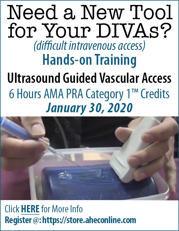
Need a new tool for your DIVAs?
If so, we got hands-on training available!
UGVA class trains in
Difficult Intra Venous Access studies. Participants attend our training to get hands-on scanning with phantoms where the ultrasound faculty teach essential skills and techniques to perform exams that make a difference in patient outcomes by demonstrating deep vein thrombosis or need for an intervention that can be done under ultrasound guidance. Our UGVA class offers not only the skills training but CME for physicians and CE for nurses.
|
|
|
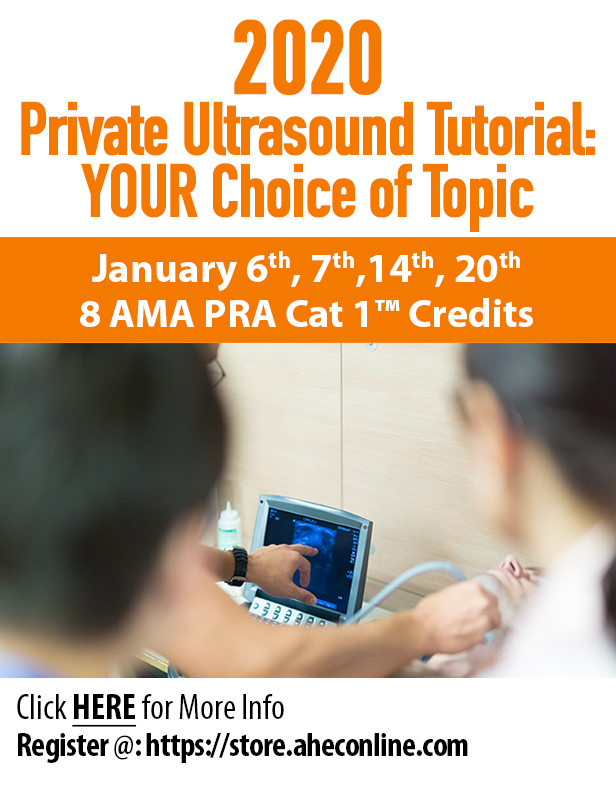
Private Ultrasound Tutorial = Convience!
Since the first ED ultrasound class in 1993, AHEC has been teaching what physicians want to learn to benefit their patients and their practice.
If you can’t make a scheduled class or want to customize your training then a Private Ultrasound Tutorial is right up your alley! We can design courses according to your practice requirements and/or skill level.
To see what upcoming dates are available for 2019. Click here:
|
|
|
Ultrasound Tip of the Month
|
|
|
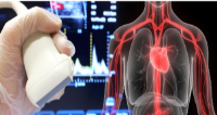
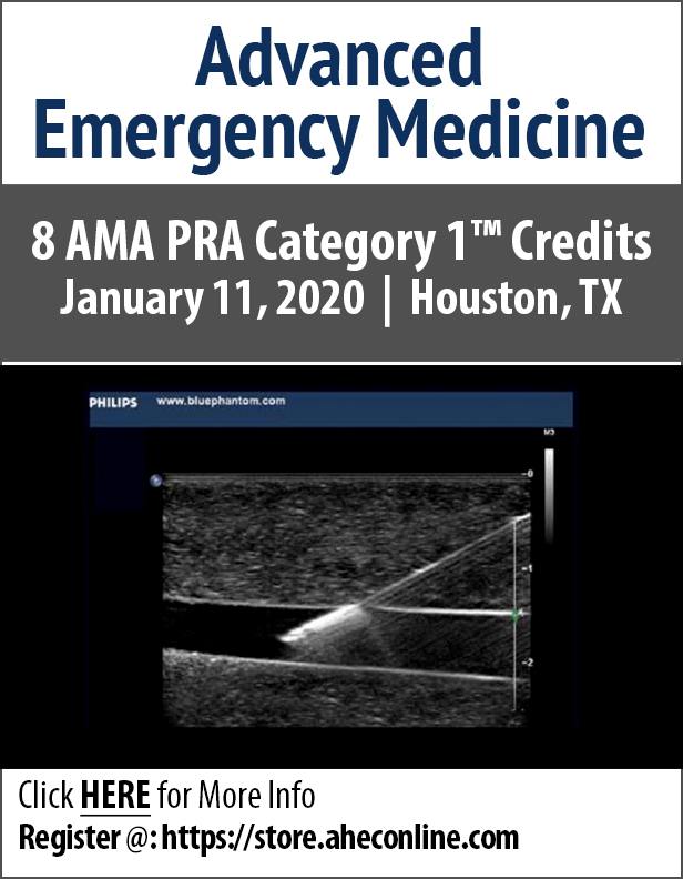
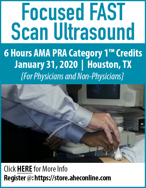
10 structures or spaces are typically imaged via 4 windows in a FAST examination; other views may be included and other structures evaluated. The windows and what is evaluated include
- Cardiac (most often subxiphoid, but other views may be obtained):
- Pericardium
- Heart chambers, especially the right ventricle
- Right Upper Quadrant (RUQ):
- Morrison's Pouch (hepatorenal recess)
- Liver tip (right paracolic gutter)
- Lower right thorax
- Left Upper Quadrant (LUQ):
- Subphrenic space
- Splenoral recess
- Spleen tip (left paracolic gutter)
- Lower left thorax
- Pelvic:
- Rectovesical pouch (male patients)
- In female patients, rectouterine/pouch of Douglas
|
|
|
|
|
|